Structures
Structural Data for proteins and associated models and simulations
Structures on this page are grouped in tiers:
- Viral VS Host
- Active interest VS low interest in drug discovery
- Therapeutic Modality (Target)
- Structure Rating
- Model Ratings
Because of the sorting, structures may appear in multiple groups under the first three categories, but they will all link out to the same locations.
Many structural annotations and reviews come from the Coronavirus Structural Task Force.
Data classification:- Structures: Data defining structures determined by experimental methods and referenced via a unique identifier such as a PDB ID.
- Models: Derived, integrated, or refined structures from multiple data sources prepared for different computational tasks.
- Targets: The biological mechanisms and functions which can be exploited to reduce, prevent, or treat the virus.
- Proteins: The biological proteins associated with the SARS-CoV-2 virus and host.
- Simulations: The datasets produced as a result of applying the models to different scientific techniques.
- Unpublished data with no preprints have no indicator
- Data in a preprint or submitted for publication are given this
 marker. If the preprint is available, it will always show and should work as a link
 - Data published in a paper or accepted are given this
 marker (i.e. has been approved by formal peer review) and should work as links.
Quick Navigation
3CLpro ACE2 BoAT1 E protein Fc receptor Furin Helicase IL6R M protein Macrodomain N protein NSP1 NSP10 NSP11 NSP14 NSP15 NSP16 NSP2 NSP4 NSP6 NSP7 NSP8 NSP9 ORF10 ORF3a ORF6 ORF7a ORF7b ORF8 PD-1 PLpro RdRP TMPRSS2 fusion core p38 spike virion
Structures of Virion Particle
---Structures of Viral Spike Proteins
Viral Spike Fusion Core
Inhibition of formation of the viral fusion core
Structure of Post Fusion Core of 2019-NCoV S2 Subunit (6LXT)
Additional: Medium-resolution structure of post-fusion core of SARS-CoV-2 S2 subunit

Structure Data and Links: PDBID: 6lxt View 3D Read the Paper
Experimental Method: X-Ray Diffraction | Resolution: 2.9 Å
Top Two Models:
Coarse Grained Post Fusion Core of 2019-NCoV S2 Subunit (PDB 6LXT)
Known Target Modalities: Inhibition of Formation of the Viral Fusion Core
Related Proteins: fusion core HR1 HR2
Top Simulations:[See All]
---
SARS-CoV-2 Spike (S) glycoprotein
Host immune response
Mapping Neutralizing and Immunodominant Sites on the SARS-CoV-2 Spike Receptor-Binding Domain by Structure-Guided High-Resolution Serology (7JX3)
Additional: Crystal structure of SARS-CoV-2 spike receptor-binding domain bound with monoclonal antibody S309

Structure Data and Links: PDBID: 7jx3 View 3D Read the Paper
Experimental Method: X-Ray Diffraction | Resolution: 2.65 Å
Top Two Models:
SARS-CoV-2 Spike Receptor-Binding Domain Bound With S309: ISOLDE Refined Model With N343 Glycan
Known Target Modalities: Blocking SARS-CoV-2 Spike Protein Binding to Human ACE2 Receptor Host Immune Response
Related Proteins: spike RBD
Top Simulations:[See All]
Antibodies to the SARS-CoV-2 Receptor-Binding Domain That Maximize Breadth and Resistance to Viral Escape (7M7W)
Additional: Crystal structure of SARS-CoV-2 spike receptor-binding domain bound with monoclonal antibody S2H97

Structure Data and Links: PDBID: 7m7w View 3D Read the Paper
Experimental Method: X-Ray Diffraction | Resolution: 2.65 Å
Top Two Models:
SARS-CoV-2 Spike Receptor-Binding Domain Bound With S2H97: ISOLDE Refined Model With N343 Glycan
Known Target Modalities: Blocking SARS-CoV-2 Spike Protein Binding to Human ACE2 Receptor Host Immune Response
Related Proteins: spike RBD
Top Simulations:[See All]
Folding@home simulations of the SARS-CoV-2 spike RBD bound to monoclonal antibody S2H97
Blocking SARS-CoV-2 Spike protein binding to human ACE2 receptor
Crystal Structure of SARS-CoV-2 Spike Receptor-Binding Domain Bound With ACE2 (6M0J)

Structure Data and Links: PDBID: 6m0j View 3D Read the Paper
Experimental Method: X-Ray Diffraction | Resolution: 2.45 Å
Top Two Models:
SARS-CoV-2 Spike Receptor-Binding Domain Bound With ACE2
SARS-CoV-2 Spike Receptor-Binding Domain Bound With ACE2: ISOLDE Refined Model
Known Target Modalities: Blocking SARS-CoV-2 Spike Protein Binding to Human ACE2 Receptor
Related Proteins: spike RBD ACE2
Top Simulations:[See All]
Folding@home simulations of the SARS-CoV-2 spike RBD with N501Y mutation bound to human ACE2
Folding@home simulations of the apo SARS-CoV-2 spike RBD (with glycosylation)
SARS-CoV-2 Spike Ectodomain Structure (Open State) (6VYB)
Additional: Low-resolution cryo-EM structure of the SARS-CoV-2 ectodomain in the open state.
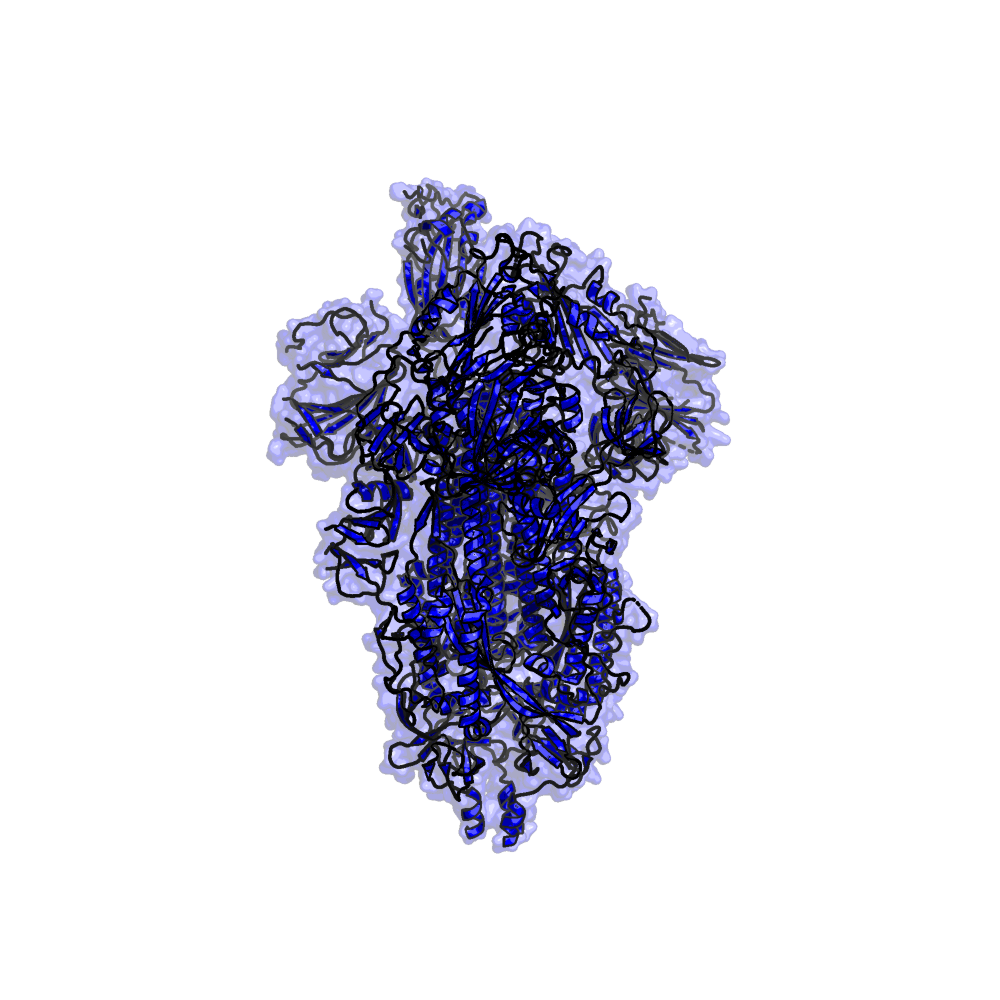
Structure Data and Links: PDBID: 6vyb View 3D Read the Paper
Experimental Method: Electron Microscopy | Resolution: 3.2 Å
Top Two Models:
Trimeric SARS-CoV-2 Spike Glycoprotein (1Up State) With and Without Simulation Box
Spike Protein in Complex With Human ACE2
Known Target Modalities: Blocking SARS-CoV-2 Spike Protein Binding to Human ACE2 Receptor
Related Proteins: spike
Top Simulations:[See All]
Cluster ensemble of 2UP like conformations
Prefusion 2019-NCoV Spike Glycoprotein With a Single Receptor-Binding Domain Up (6VSB)

Structure Data and Links: PDBID: 6VSB View 3D Read the Paper
Experimental Method: Electron Microscopy | Resolution: 3.46 Å
Top Two Models:
Furin Bound to Delta Variant SARS-CoV-2 Spike in One RBD Up State
Spike_full-Length_open_amarolab
Known Target Modalities: Blocking SARS-CoV-2 Spike Protein Binding to Human ACE2 Receptor
Related Proteins: spike RBD
Top Simulations:[See All]
Riken CPR TMS, MD1_Up trajectory
Riken CPR TMS, MD2_Up trajectory
Structure of the SARS-CoV-2 Spike Glycoprotein (Closed State) (6VXX)

Structure Data and Links: PDBID: 6VXX View 3D Read the Paper
Experimental Method: Electron Microscopy | Resolution: 2.8 Å
Top Two Models:
SARS-CoV-2 Trimeric Spike Protein Binding to FDA Approved or Investigational Drug Molecules
Improved Trimeric SARS-CoV-2 Spike Glycoprotein (Closed State) in Aqueous Solution
Known Target Modalities: Blocking SARS-CoV-2 Spike Protein Binding to Human ACE2 Receptor
Related Proteins: spike
Top Simulations:[See All]
Trajectories of full-length SPIKE protein in the Closed state.
Cluster ensemble of Intermediate 3a
Mapping Neutralizing and Immunodominant Sites on the SARS-CoV-2 Spike Receptor-Binding Domain by Structure-Guided High-Resolution Serology (7JX3)
Additional: Crystal structure of SARS-CoV-2 spike receptor-binding domain bound with monoclonal antibody S309
Structure Data and Links: PDBID: 7jx3 View 3D Read the Paper
Experimental Method: X-Ray Diffraction | Resolution: 2.65 Å
Top Two Models:
SARS-CoV-2 Spike Receptor-Binding Domain Bound With S309: ISOLDE Refined Model With N343 Glycan
Known Target Modalities: Blocking SARS-CoV-2 Spike Protein Binding to Human ACE2 Receptor Host Immune Response
Related Proteins: spike RBD
Top Simulations:[See All]
Crystal Structure of SARS-CoV-2 Receptor Binding Domain in Complex With Human Antibody CR3022 (6W41)
Additional: Low resolution X-ray structure of human antibody CR3022 bound to the receptor binding domain (RBD) of the spike protein.

Structure Data and Links: PDBID: 6w41 View 3D Read the Paper
Experimental Method: X-Ray Diffraction | Resolution: 3.084 Å
Top Two Models:
Coarse Grained RBD in Complex With Human Antibody CR3022 (PDB 6W41)
Known Target Modalities: Blocking SARS-CoV-2 Spike Protein Binding to Human ACE2 Receptor
Related Proteins: spike RBD
Top Simulations:[See All]
SIRAH-CoV2 initiative - S1 Receptor Binding Domain in complex with human antibody CR3022
Structure of SARS Coronavirus Spike Receptor-Binding Domain Complexed With Its Receptor (2AJF)
Additional: High-resolution cryo-EM structure of the full-length SARS-CoV-1 spike protein bound to human host cell receptor ACE2.
Structure Data and Links: PDBID: 2AJF View 3D Read the Paper
Experimental Method: X-Ray Diffraction | Resolution: 2.9 Å
Top Two Models:
Coarse Grained SPIKE Protein (PDB 2AJF)
Known Target Modalities: Blocking SARS-CoV-2 Spike Protein Binding to Human ACE2 Receptor
Related Proteins: spike RBD ACE2
Top Simulations:[See All]
Simulations of SARS-CoV and SARS-CoV-2 RBD with ACE2
A 10 µs simulation of a SARS-CoV-1 and SARS-CoV-2 chimera-ACE2 complex in aqueous solution
Antibodies to the SARS-CoV-2 Receptor-Binding Domain That Maximize Breadth and Resistance to Viral Escape (7M7W)
Additional: Crystal structure of SARS-CoV-2 spike receptor-binding domain bound with monoclonal antibody S2H97
Structure Data and Links: PDBID: 7m7w View 3D Read the Paper
Experimental Method: X-Ray Diffraction | Resolution: 2.65 Å
Top Two Models:
SARS-CoV-2 Spike Receptor-Binding Domain Bound With S2H97: ISOLDE Refined Model With N343 Glycan
Known Target Modalities: Blocking SARS-CoV-2 Spike Protein Binding to Human ACE2 Receptor Host Immune Response
Related Proteins: spike RBD
Top Simulations:[See All]
Folding@home simulations of the SARS-CoV-2 spike RBD bound to monoclonal antibody S2H97
Cryo-EM Structure of the SARS-CoV-2 Spike Glycoprotein Bound to Fab 2-4 (6XEY)

Structure Data and Links: PDBID: 6XEY View 3D Read the Paper
Experimental Method: Electron Microscopy | Resolution: 3.25 Å
Top Two Models:
---
Known Target Modalities: Blocking SARS-CoV-2 Spike Protein Binding to Human ACE2 Receptor
Related Proteins: spike RBD
Top Simulations:[See All]
SIRAH-CoV2 initiative - RBD triple glycosylated at Asn331, 343, and 481
The 2019-NCoV RBD/ACE2-B0AT1 Complex (6M17)
Additional: High-resolution cryo-EM structure of the full-length SARS-CoV-2 spike protein bound to human host cell receptor ACE2.
Structure Data and Links: PDBID: 6m17 View 3D Read the Paper
Experimental Method: Electron Microscopy | Resolution: 2.9 Å
Top Two Models:
Coarse Grained RBD/ACE2-B0AT1 Complex (PDB 6M17)
Spike Protein in Complex With Human ACE2
Known Target Modalities: Blocking SARS-CoV-2 Spike Protein Binding to Human ACE2 Receptor
Related Proteins: spike RBD ACE2 BoAT1
Top Simulations:[See All]
DESRES-ANTON-10905033 10 µs simulation of the SARS-CoV-2-ACE2 complex in aqueous solution
Simulations of SARS-CoV and SARS-CoV-2 RBD with ACE2 possessing different patterns of glycosylation
Crystal Structure of Post Fusion Core of 2019-NCoV S2 Subunit (6M1V)

Structure Data and Links: PDBID: 6M1V View 3D Read the Paper
Experimental Method: X-Ray Diffraction | Resolution: 1.5 Å
Top Two Models:
---
Known Target Modalities: Blocking SARS-CoV-2 Spike Protein Binding to Human ACE2 Receptor
Related Proteins: spike S2
Top Simulations:[See All]
SIRAH-CoV2 initiative - S2 Spike core fragment in postfusion state
Structures of Viral Protease, Polymerase, and Nonstructured Proteins
SARS-CoV-2 main protease (3CLpro or NSP5)
3CLpro / Mpro activity
Crystal Structure (Monoclinic Form) of the Complex Resulting From the Reaction Between SARS-CoV-2 (2019-NCoV) Main Protease and Tert-Butyl (1-((S)-1-(((S)-4-(Benzylamino)-3,4-Dioxo-1-((S)-2-Oxopyrrolidin-3-Yl)butan-2-Yl)amino)-3-Cyclopropyl-1-Oxopropan-2-Yl)-2-Oxo-1,2-Dihydropyridin-3-Yl)carbamate (Alpha-Ketoamide 13b) (6Y2F)
Additional: Crystal structure of SARS-CoV-2 main protease provides a basis for drug repurposing studies

Structure Data and Links: PDBID: 6Y2F View 3D Read the Paper
Experimental Method: X-Ray Diffraction | Resolution: 1.95 Å
Top Two Models:
Truncated Mpro Based on 6Y2F and Shape-Compliant 3D Conformers
Truncated Mpro Based on 6Y2F and Pharmacophore-Compliant 3D Conformers
Known Target Modalities: 3CLpro / Mpro Activity
Related Proteins: 3CLpro
Top Simulations:[See All]
HADDOCK docking of approved Drugbank set against Mpro with a pharmacophore shape model
HADDOCK docking of approved Drugbank set against Mpro with a geometric shape model
6LZE
Additional: X-Ray crystal structure of SARS-Cov-2 MPro in complex with 11a
Structure Data and Links: PDBID: View 3D
Top Two Models:
---
Known Target Modalities: 3CLpro / Mpro Activity
Related Proteins: 3CLpro
Top Simulations:[See All]
---
Crystal Structure (Orthorhombic Form) of the Complex Resulting From the Reaction Between SARS-CoV-2 (2019-NCoV) Main Protease and Tert-Butyl (1-((S)-1-(((S)-4-(Benzylamino)-3,4-Dioxo-1-((S)-2-Oxopyrrolidin-3-Yl)butan-2-Yl)amino)-3-Cyclopropyl-1-Oxopropan-2-Yl)-2-Oxo-1,2-Dihydropyridin-3-Yl)carbamate (Alpha-Ketoamide 13b) (6Y2G)
Additional: Crystal structure of SARS-CoV-2 main protease provides a basis for design of improved alpha-ketoamide inhibitors

Structure Data and Links: PDBID: 6Y2G View 3D Read the Paper
Experimental Method: X-Ray Diffraction | Resolution: 2.2 Å
Top Two Models:
Coarse Grained 6y2g (PDB 6Y2G)
Known Target Modalities: 3CLpro / Mpro Activity
Related Proteins: 3CLpro
Top Simulations:[See All]
---
SARS-CoV-2 Main Protease With Unliganded Active Site (2019-NCoV, Coronavirus Disease 2019, COVID-19) (6Y84)
Structure Data and Links: PDBID: 6Y84 View 3D To Be Published
Experimental Method: X-Ray Diffraction | Resolution: 1.39 Å
Top Two Models:
3CLpro Prepared for Simulation in a 120 Cubic a Box for Long Continuous Trajectory
Known Target Modalities: 3CLpro / Mpro Activity
Related Proteins: 3CLpro
Top Simulations:[See All]
DESRES 100 µs MD of 3CLpro, no water or ions
DESRES 100 µs MD of 3CLpro, All Atom
The Crystal Structure of COVID-19 Main Protease in Complex With an Inhibitor N3 (6LU7)

Structure Data and Links: PDBID: 6LU7 View 3D Read the Paper
Experimental Method: X-Ray Diffraction | Resolution: 2.16 Å
Top Two Models:
SARS-CoV-2 Main Protease (Apo, Monomer) for Folding@home Simulations
SARS-CoV-2 Dimeric Main Protease Without Ligand Based on PDB 6LU7
Known Target Modalities: 3CLpro / Mpro Activity
Related Proteins: 3CLpro
Top Simulations:[See All]
Riken BDR 10 Microsecond Trajectory Protein Snapshot every 1ns
SIRAH-CoV2 initiative - Main Protease
The Crystal Structure of COVID-19 Main Protease in Complex With Carmofur (7BUY)

Structure Data and Links: PDBID: 7BUY View 3D Read the Paper
Experimental Method: X-Ray Diffraction | Resolution: 1.6 Å
Top Two Models:
Known Target Modalities: Inhibition of Viral Polymerases 3CLpro / Mpro Activity
Related Proteins: 3CLpro
Top Simulations:[See All]
---
PanDDA Analysis Group Deposition of Ground-State Model of SARS-CoV-2 Main Protease Screened Against DSI Poised (Enamine), Fraglites and Peplites (Newcastle University), Mini Frags (Astex), York 3D (York University), Electrophile Cysteine Covalent (Weizman Institute) Fragment Libraries (5R8T)
Additional: PanDDA analysis group deposition of ground-state model of SARS-CoV-2 main protease fragment screening

Structure Data and Links: PDBID: 5R8T View 3D To Be Published
Experimental Method: X-Ray Diffraction | Resolution: 1.27 Å
Top Two Models:
---
Known Target Modalities: 3CLpro / Mpro Activity
Related Proteins: 3CLpro
Top Simulations:[See All]
---
Inhibition of viral polymerases
The Crystal Structure of COVID-19 Main Protease in Complex With Carmofur (7BUY)
Structure Data and Links: PDBID: 7BUY View 3D Read the Paper
Experimental Method: X-Ray Diffraction | Resolution: 1.6 Å
Top Two Models:
Known Target Modalities: Inhibition of Viral Polymerases 3CLpro / Mpro Activity
Related Proteins: 3CLpro
Top Simulations:[See All]
---
SARS-CoV-2 Macrodomain (NSP3)
SARS-CoV-2 Papain-like protease (NSP3)
3CLpro / Mpro activity
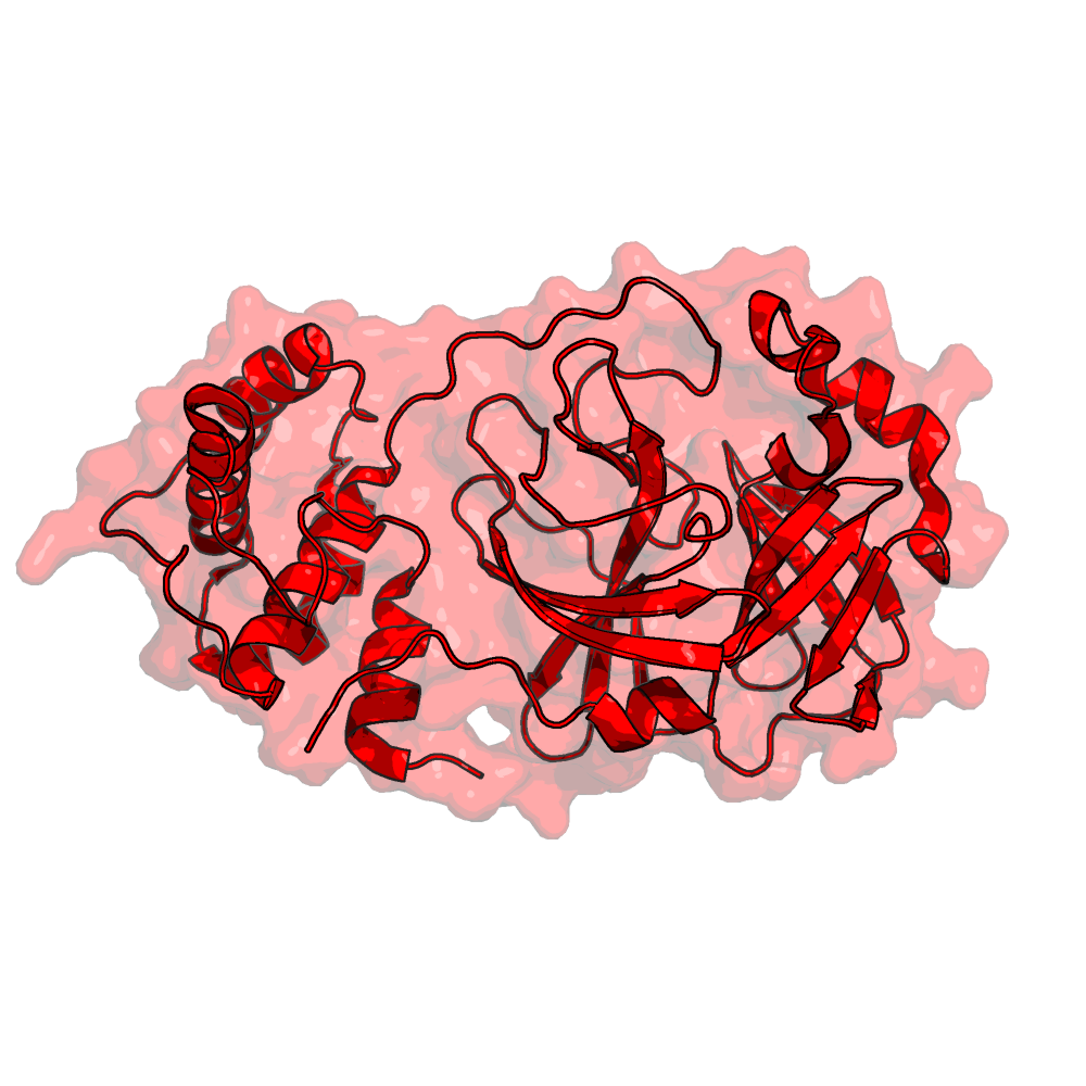
Crystal Structure of ADP Ribose Phosphatase of NSP3 From SARS CoV-2 in the Complex With ADP Ribose (6W02)
Additional: Crystal Structure of ADP ribose phosphatase of PLpro from SARS CoV-2 in the complex with ADP ribose

Structure Data and Links: PDBID: 6W02 View 3D Read the Paper
Experimental Method: X-Ray Diffraction | Resolution: 1.5 Å
Top Two Models:
---
Known Target Modalities: 3CLpro / Mpro Activity
Related Proteins: PLpro
Top Simulations:[See All]
SIRAH-CoV2 initiative - Apo ADP-ribose phosphatase of NSP3
Folding@home simulations of nsp3 macrodomain
Inhibition of PLpro protease activity
The Crystal Structure of Papain-Like Protease of SARS CoV-2 (6W9C)

Structure Data and Links: PDBID: 6w9c View 3D Read the Paper
Experimental Method: X-Ray Diffraction | Resolution: 2.7 Å
Top Two Models:
SARS-CoV-2 PLpro: ISOLDE Refined Model
SARS-CoV-2 Ligand-Bound (3k Ligand Was Docked to Protein Conformation From 6W9C Ligand-Free MD)
Known Target Modalities: Inhibition of PLpro Protease Activity
Related Proteins: PLpro
Top Simulations:[See All]
3k bound SARS-CoV-2 PLPro (3k docked to frame from trajectory of PDB 6W9C C-chain)
SIRAH-CoV2 initiative - Papain-like Protease
The Crystal Structure of Papain-Like Protease of SARS CoV-2 , C111S Mutant (6WRH)

Structure Data and Links: PDBID: 6wrh View 3D Read the Paper
Experimental Method: X-Ray Diffraction | Resolution: 1.6 Å
Top Two Models:
SARS-CoV-2 Ligand-Free (PDB 6WRH)
Known Target Modalities: Inhibition of PLpro Protease Activity
Related Proteins: PLpro
Top Simulations:[See All]
Apo SARS-CoV-2 PLPro (from PDB 6WRH C-chain)
X-Ray Structural and Biological Evaluation of a Series of Potent and Highly Selective Inhibitors of Human Coronavirus Papain-Like Proteases (4OW0)

Structure Data and Links: PDBID: 4ow0 View 3D Read the Paper
Experimental Method: X-Ray Diffraction | Resolution: 2.1 Å
Top Two Models:
Coarse Grained Inhibitors of Human Coronavirus Papain-Like Proteases (PDB 4OW0 Ligands Removed)
SARS-CoV-1 Ligand-Free (PDB 4OW0 Ligand Removed)
Known Target Modalities: Inhibition of PLpro Protease Activity
Related Proteins: PLpro
Top Simulations:[See All]
Inhibition of viral polymerases
SARS Coronavirus Papain-Like Protease: Structure of a Viral Deubiquitinating Enzyme (2FE8)
Structure Data and Links: PDBID: 2FE8 View 3D Read the Paper
Experimental Method: X-Ray Diffraction | Resolution: 1.85 Å
Top Two Models:
Coarse Grained Papain-Like Protease (PDB 2FE8)
Known Target Modalities: Inhibition of Viral Polymerases
Related Proteins: PLpro
Top Simulations:[See All]
---
SARS-CoV-2 RNA Polymerase (NSP12)
Inhibition of viral polymerases
Cryo-EM Structure of the Apo Nsp12-Nsp7-Nsp8 Complex (7BV1)

Structure Data and Links: PDBID: 7BV1 View 3D Read the Paper
Experimental Method: Electron Microscopy | Resolution: 2.8 Å
Top Two Models:
Coarse Grained Apo NSP7, NSP8 Molecules (PDB 7BV1)
SARS-CoV-2 Apo-RdRp Complex (Nsp12+2*nsp8+nsp7) Model for MD Simulations
Known Target Modalities: Inhibition of Viral Polymerases
Related Proteins: RdRP NSP7 NSP8
Top Simulations:[See All]
Gromacs 300 ns MD of SARS-CoV-2 apo-RdRp model, All Atom model
SARS-CoV-2 RNA-Dependent RNA Polymerase in Complex With Cofactors in Reduced Condition (7BTF)

Structure Data and Links: PDBID: 7BTF View 3D Read the Paper
Experimental Method: Electron Microscopy | Resolution: 2.95 Å
Top Two Models:
SARS-CoV-2 RdRp Complex (Nsp12+2*nsp8+nsp7) + RNA Template-Primer + ATP Model for MD Simulations
SARS-CoV-2 RdRP (NSP12) in Complex With Cofactors NSP7 and NSP8: ISOLDE Refined Model
Known Target Modalities: Inhibition of Viral Polymerases
Related Proteins: RdRP NSP7 NSP8
Top Simulations:[See All]
Gromacs 300 ns MD of SARS-CoV-2 apo-RdRp model, All Atom model
SARS-Cov-2 RNA-Dependent RNA Polymerase in Complex With Cofactors (6M71)
Additional: High-resolution cyro-EM structure of the SARS-CoV-2 NSP7-8 complex with RdRP
Structure Data and Links: PDBID: 6m71 View 3D Read the Paper
Experimental Method: Electron Microscopy | Resolution: 2.9 Å
Top Two Models:
SARS-CoV-2 Apo-RdRp Complex (Nsp12+2*nsp8+nsp7) Model for MD Simulations
Known Target Modalities: Inhibition of Viral Polymerases
Related Proteins: NSP7 NSP8 RdRP
Top Simulations:[See All]
The Nsp12-Nsp7-Nsp8 Complex Bound to the Template-Primer RNA and Triphosphate Form of Remdesivir(RTP) (7BV2)

Structure Data and Links: PDBID: 7BV2 View 3D Read the Paper
Experimental Method: Electron Microscopy | Resolution: 2.5 Å
Top Two Models:
Docking-Based Repurposing Study of Approved Drugs Against Truncated RdRp
Known Target Modalities: Inhibition of Viral Polymerases
Related Proteins: NSP7 NSP8 RdRP
Top Simulations:[See All]
Gromacs 100 ns MD of SARS-CoV-2 RdRp + RNA template-primer + ATP model, All Atom model
HADDOCK docking of approved Drugbank set against RdRp
Helicase coronavirus nonstructural protein 13 (NSP13)
Inhibition of viral polymerases
Crystal Structure of the SARS-CoV-2 Helicase at 1.94 Angstrom Resolution (6ZSL)

Structure Data and Links: PDBID: 6ZSL View 3D Read the Paper
Experimental Method: X-Ray Diffraction | Resolution: 1.94 Å
Top Two Models:
---
Known Target Modalities: Inhibition of Viral Polymerases
Related Proteins: Helicase
Top Simulations:[See All]
SIRAH-CoV2 initiative - Helicase
SARS CoV-2 Spike Protein, Closed Conformation, C3 Symmetry (6ZB5)

Structure Data and Links: PDBID: 6ZB5 View 3D Read the Paper
Experimental Method: Electron Microscopy | Resolution: 2.85 Å
Top Two Models:
---
Known Target Modalities: Inhibition of Viral Polymerases
Related Proteins: Helicase
Top Simulations:[See All]
Coronavirus nonstructural protein 1
Coronavirus nonstructural protein 10
Inhibition of viral polymerases
1.80 Angstrom Resolution Crystal Structure of NSP16 - NSP10 Complex From SARS-CoV-2 (6W4H)

Structure Data and Links: PDBID: 6W4H View 3D Read the Paper
Experimental Method: X-Ray Diffraction | Resolution: 1.8 Å
Top Two Models:
---
Known Target Modalities: Inhibition of Viral Polymerases
Related Proteins: NSP16 NSP10
Top Simulations:[See All]
SIRAH-CoV2 initiative - NSP16 - NSP10 Complex
Folding@home simulations of nsp10
Coronavirus nonstructural protein 11
Coronavirus nonstructural protein 14
Coronavirus nonstructural protein 15
Inhibition of viral polymerases
The Crystal Structure of COVID-19 Main Protease in Apo Form (6M03)

Structure Data and Links: PDBID: 6M03 View 3D Read the Paper
Experimental Method: X-Ray Diffraction | Resolution: 2 Å
Top Two Models:
Coarse Grained COVID-19 Main Protease in Apo Form (PDB 6M03)
Docked Structures of a Small Molecule Inhibitor (X77) to SARS-CoV-2 Mpro
Known Target Modalities: Inhibition of Viral Polymerases
Related Proteins: NSP15
Top Simulations:[See All]
---
The 1.9 a Crystal Structure of NSP15 Endoribonuclease From SARS CoV-2 in the Complex With a Citrate (6W01)

Structure Data and Links: PDBID: 6W01 View 3D Read the Paper
Experimental Method: X-Ray Diffraction | Resolution: 1.9 Å
Top Two Models:
---
Known Target Modalities: Inhibition of Viral Polymerases
Related Proteins: NSP15
Top Simulations:[See All]
SIRAH-CoV2 initiative - NSP15 Endonuclease
Coronavirus nonstructural protein 16
Inhibition of viral polymerases
1.80 Angstrom Resolution Crystal Structure of NSP16 - NSP10 Complex From SARS-CoV-2 (6W4H)
Structure Data and Links: PDBID: 6W4H View 3D Read the Paper
Experimental Method: X-Ray Diffraction | Resolution: 1.8 Å
Top Two Models:
---
Known Target Modalities: Inhibition of Viral Polymerases
Related Proteins: NSP16 NSP10
Top Simulations:[See All]
Folding@home simulations of nsp10
SIRAH-CoV2 initiative - NSP16 - NSP10 Complex
Coronavirus nonstructural protein 2
Coronavirus nonstructural protein 4
Coronavirus nonstructural protein 6
Coronavirus nonstructural protein 7
Inhibition of viral polymerases
Crystal Structure of the Co-Factor Complex of NSP7 and the C-Terminal Domain of NSP8 From SARS CoV-2 (6WIQ)

Structure Data and Links: PDBID: 6WIQ View 3D Read the Paper
Experimental Method: X-Ray Diffraction | Resolution: 2.85 Å
Top Two Models:
---
Known Target Modalities: Inhibition of Viral Polymerases
Related Proteins: NSP7 NSP8
Top Simulations:[See All]
SIRAH-CoV2 initiative - Co-factor complex of NSP7 and the C-terminal domain of NSP8
Crystal Structure of the Nsp7-Nsp8 Complex of SARS-CoV-2 (6YHU)

Structure Data and Links: PDBID: 6YHU View 3D Read the Paper
Experimental Method: X-Ray Diffraction | Resolution: 2 Å
Top Two Models:
---
Known Target Modalities: Inhibition of Viral Polymerases
Related Proteins: NSP7 NSP8
Top Simulations:[See All]
SIRAH-CoV2 initiative - NSP7-NSP8 complex
SARS-CoV-2 RNA-Dependent RNA Polymerase in Complex With Cofactors in Reduced Condition (7BTF)
Structure Data and Links: PDBID: 7BTF View 3D Read the Paper
Experimental Method: Electron Microscopy | Resolution: 2.95 Å
Top Two Models:
SARS-CoV-2 Apo-RdRp Complex (Nsp12+2*nsp8+nsp7) Model for MD Simulations
SARS-CoV-2 RdRp Complex (Nsp12+2*nsp8+nsp7) + RNA Template-Primer + ATP Model for MD Simulations
Known Target Modalities: Inhibition of Viral Polymerases
Related Proteins: RdRP NSP7 NSP8
Top Simulations:[See All]
SIRAH-CoV2 initiative - RNA-dependent RNA polymerase in complex with cofactors NSP7 and NSP8
Gromacs 100 ns MD of SARS-CoV-2 RdRp + RNA template-primer + ATP model, All Atom model
Cryo-EM Structure of the Apo Nsp12-Nsp7-Nsp8 Complex (7BV1)
Structure Data and Links: PDBID: 7BV1 View 3D Read the Paper
Experimental Method: Electron Microscopy | Resolution: 2.8 Å
Top Two Models:
Coarse Grained Apo NSP7, NSP8 Molecules (PDB 7BV1)
SARS-CoV-2 Apo-RdRp Complex (Nsp12+2*nsp8+nsp7) Model for MD Simulations
Known Target Modalities: Inhibition of Viral Polymerases
Related Proteins: RdRP NSP7 NSP8
Top Simulations:[See All]
Gromacs 300 ns MD of SARS-CoV-2 apo-RdRp model, All Atom model
SARS-Cov-2 RNA-Dependent RNA Polymerase in Complex With Cofactors (6M71)
Additional: High-resolution cyro-EM structure of the SARS-CoV-2 NSP7-8 complex with RdRP
Structure Data and Links: PDBID: 6m71 View 3D Read the Paper
Experimental Method: Electron Microscopy | Resolution: 2.9 Å
Top Two Models:
SARS-CoV-2 Apo-RdRp Complex (Nsp12+2*nsp8+nsp7) Model for MD Simulations
Known Target Modalities: Inhibition of Viral Polymerases
Related Proteins: NSP7 NSP8 RdRP
Top Simulations:[See All]
Gromacs 300 ns MD of SARS-CoV-2 apo-RdRp model, All Atom model
The Nsp12-Nsp7-Nsp8 Complex Bound to the Template-Primer RNA and Triphosphate Form of Remdesivir(RTP) (7BV2)
Structure Data and Links: PDBID: 7BV2 View 3D Read the Paper
Experimental Method: Electron Microscopy | Resolution: 2.5 Å
Top Two Models:
Docking-Based Repurposing Study of Approved Drugs Against Truncated RdRp
Known Target Modalities: Inhibition of Viral Polymerases
Related Proteins: NSP7 NSP8 RdRP
Top Simulations:[See All]
Gromacs 100 ns MD of SARS-CoV-2 RdRp + RNA template-primer + ATP model, All Atom model
Coronavirus nonstructural protein 8
Inhibition of viral polymerases
SARS-CoV-2 RNA-Dependent RNA Polymerase in Complex With Cofactors in Reduced Condition (7BTF)
Structure Data and Links: PDBID: 7BTF View 3D Read the Paper
Experimental Method: Electron Microscopy | Resolution: 2.95 Å
Top Two Models:
SARS-CoV-2 Apo-RdRp Complex (Nsp12+2*nsp8+nsp7) Model for MD Simulations
SARS-CoV-2 RdRp Complex (Nsp12+2*nsp8+nsp7) + RNA Template-Primer + ATP Model for MD Simulations
Known Target Modalities: Inhibition of Viral Polymerases
Related Proteins: RdRP NSP7 NSP8
Top Simulations:[See All]
Gromacs 300 ns MD of SARS-CoV-2 apo-RdRp model, All Atom model
SIRAH-CoV2 initiative - RNA-dependent RNA polymerase in complex with cofactors NSP7 and NSP8
Cryo-EM Structure of the Apo Nsp12-Nsp7-Nsp8 Complex (7BV1)
Structure Data and Links: PDBID: 7BV1 View 3D Read the Paper
Experimental Method: Electron Microscopy | Resolution: 2.8 Å
Top Two Models:
SARS-CoV-2 Apo-RdRp Complex (Nsp12+2*nsp8+nsp7) Model for MD Simulations
Coarse Grained Apo NSP7, NSP8 Molecules (PDB 7BV1)
Known Target Modalities: Inhibition of Viral Polymerases
Related Proteins: RdRP NSP7 NSP8
Top Simulations:[See All]
Gromacs 300 ns MD of SARS-CoV-2 apo-RdRp model, All Atom model
Crystal Structure of the Co-Factor Complex of NSP7 and the C-Terminal Domain of NSP8 From SARS CoV-2 (6WIQ)
Structure Data and Links: PDBID: 6WIQ View 3D Read the Paper
Experimental Method: X-Ray Diffraction | Resolution: 2.85 Å
Top Two Models:
---
Known Target Modalities: Inhibition of Viral Polymerases
Related Proteins: NSP7 NSP8
Top Simulations:[See All]
SIRAH-CoV2 initiative - Co-factor complex of NSP7 and the C-terminal domain of NSP8
Crystal Structure of the Nsp7-Nsp8 Complex of SARS-CoV-2 (6YHU)
Structure Data and Links: PDBID: 6YHU View 3D Read the Paper
Experimental Method: X-Ray Diffraction | Resolution: 2 Å
Top Two Models:
---
Known Target Modalities: Inhibition of Viral Polymerases
Related Proteins: NSP7 NSP8
Top Simulations:[See All]
SIRAH-CoV2 initiative - NSP7-NSP8 complex
SARS-Cov-2 RNA-Dependent RNA Polymerase in Complex With Cofactors (6M71)
Additional: High-resolution cyro-EM structure of the SARS-CoV-2 NSP7-8 complex with RdRP
Structure Data and Links: PDBID: 6m71 View 3D Read the Paper
Experimental Method: Electron Microscopy | Resolution: 2.9 Å
Top Two Models:
SARS-CoV-2 RdRP (NSP12) in Complex With NSP7 and Two Copies of NSP8: ISOLDE Refined Model
Known Target Modalities: Inhibition of Viral Polymerases
Related Proteins: NSP7 NSP8 RdRP
Top Simulations:[See All]
Gromacs 100 ns MD of SARS-CoV-2 RdRp + RNA template-primer + ATP model, All Atom model
The Nsp12-Nsp7-Nsp8 Complex Bound to the Template-Primer RNA and Triphosphate Form of Remdesivir(RTP) (7BV2)
Structure Data and Links: PDBID: 7BV2 View 3D Read the Paper
Experimental Method: Electron Microscopy | Resolution: 2.5 Å
Top Two Models:
Docking-Based Repurposing Study of Approved Drugs Against Truncated RdRp
Known Target Modalities: Inhibition of Viral Polymerases
Related Proteins: NSP7 NSP8 RdRP
Top Simulations:[See All]
Gromacs 100 ns MD of SARS-CoV-2 RdRp + RNA template-primer + ATP model, All Atom model
Coronavirus nonstructural protein 9
Inhibition of viral polymerases
The Crystal Structure of Nsp9 RNA Binding Protein of SARS CoV-2 (6W4B)

Structure Data and Links: PDBID: 6W4B View 3D To Be Published
Experimental Method: X-Ray Diffraction | Resolution: 2.95 Å
Top Two Models:
---
Known Target Modalities: Inhibition of Viral Polymerases
Related Proteins: NSP9
Top Simulations:[See All]
SIRAH-CoV2 initiative - NSP9 RNA binding protein
Folding@home simulations of nsp9
Structures of Viral Open Reading Frame Proteins
Coronavirus Open Reading Frame 10
Coronavirus Open Reading Frame 3a
Host immune response
Cryo-EM Structure of SARS-CoV-2 ORF3a (6XDC)

Structure Data and Links: PDBID: 6XDC View 3D Read the Paper
Experimental Method: Electron Microscopy | Resolution: 2.9 Å
Top Two Models:
---
Known Target Modalities: Host Immune Response
Related Proteins: ORF3a
Top Simulations:[See All]
SIRAH-CoV2 initiative - Membrane embedded ORF3a
Coronavirus Open Reading Frame 6
Coronavirus Open Reading Frame 7a
Host immune response
STRUCTURE of the SARS-CoV-2 ORF7A ENCODED ACCESSORY PROTEIN (6W37)

Structure Data and Links: PDBID: 6W37 View 3D Read the Paper
Experimental Method: X-Ray Diffraction | Resolution: 2.9 Å
Top Two Models:
---
Known Target Modalities: Host Immune Response
Related Proteins: ORF7a
Top Simulations:[See All]
SIRAH-CoV2 initiative - ORF7A enconded accessory protein
Coronavirus Open Reading Frame 7b
Coronavirus Open Reading Frame 8
Structures of Viral Membrane Proteins
Membrane Glycoprotein
Structures of Viral Envelope Proteins
Envelope small membrane protein
Structures of Viral Nucleocapsid Proteins
Nucleoprotein
Inhibition of formation of the viral fusion core
Crystal Structure of RNA Binding Domain of Nucleocapsid Phosphoprotein From SARS Coronavirus 2 (6VYO)

Structure Data and Links: PDBID: 6VYO View 3D To Be Published
Experimental Method: X-Ray Diffraction | Resolution: 1.7 Å
Top Two Models:
---
Known Target Modalities: Inhibition of Formation of the Viral Fusion Core
Related Proteins: N protein
Top Simulations:[See All]
SIRAH-CoV2 initiative - RNA binding domain of nucleocapsid phosphoprotein
Crystal Structure of SARS-CoV-2 Nucleocapsid Protein N-Terminal RNA Binding Domain (6M3M)

Structure Data and Links: PDBID: 6M3M View 3D Read the Paper
Experimental Method: X-Ray Diffraction | Resolution: 2.7 Å
Top Two Models:
---
Known Target Modalities: Inhibition of Formation of the Viral Fusion Core
Related Proteins: N protein
Top Simulations:[See All]
SIRAH-CoV2 initiative - Nucleocapsid protein N-terminal RNA binding domain
Structures of Host Proteins
Angiotensin-converting enzyme 2 (ACE2)
Blocking SARS-CoV-2 Spike protein binding to human ACE2 receptor
The 2019-NCoV RBD/ACE2-B0AT1 Complex (6M17)
Additional: High-resolution cryo-EM structure of the full-length SARS-CoV-2 spike protein bound to human host cell receptor ACE2.
Structure Data and Links: PDBID: 6m17 View 3D Read the Paper
Experimental Method: Electron Microscopy | Resolution: 2.9 Å
Top Two Models:
Coarse Grained RBD/ACE2-B0AT1 Complex (PDB 6M17)
Spike Protein in Complex With Human ACE2
Known Target Modalities: Blocking SARS-CoV-2 Spike Protein Binding to Human ACE2 Receptor
Related Proteins: spike RBD ACE2 BoAT1
Top Simulations:[See All]
Trajectory of the Spike protein in complex with human ACE2
Simulations of SARS-CoV and SARS-CoV-2 RBD with ACE2
Crystal Structure of SARS-CoV-2 Spike Receptor-Binding Domain Bound With ACE2 (6M0J)
Structure Data and Links: PDBID: 6m0j View 3D Read the Paper
Experimental Method: X-Ray Diffraction | Resolution: 2.45 Å
Top Two Models:
SARS-CoV-2 Spike Receptor-Binding Domain Bound With ACE2: ISOLDE Refined Model
SARS-CoV-2 Spike Receptor-Binding Domain: ISOLDE Refined Model With N343 Glycan and N501Y Mutation
Known Target Modalities: Blocking SARS-CoV-2 Spike Protein Binding to Human ACE2 Receptor
Related Proteins: spike RBD ACE2
Top Simulations:[See All]
Folding@home simulations of the apo SARS-CoV-2 spike RBD (without glycosylation)
Folding@home simulations of the SARS-CoV-2 spike RBD with N501Y mutation bound to human ACE2
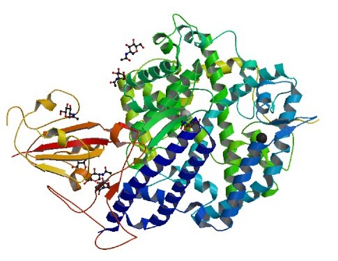
Inhibitor Bound Human Angiotensin Converting Enzyme-Related Carboxypeptidase (ACE2) (1R4L)
Structure Data and Links: PDBID: 1R4L View 3D Read the Paper
Experimental Method: X-Ray Diffraction | Resolution: 3 Å
Top Two Models:
Human ACE2 Ectodomain in Aqueous Solution (Inhibitor-Bound Closed State)
Known Target Modalities: Blocking SARS-CoV-2 Spike Protein Binding to Human ACE2 Receptor
Related Proteins: ACE2
Top Simulations:[See All]
HADDOCK docking of approved Drugbank set against human ACE2 ectodomain
DESRES-ANTON-10875754 10 µs simulation trajectory of the human ACE2 ectodomain in aqueous solution
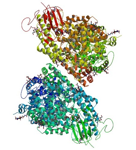
Native Human Angiotensin Converting Enzyme-Related Carboxypeptidase (ACE2) (1R42)
Structure Data and Links: PDBID: 1R42 View 3D Read the Paper
Experimental Method: X-Ray Diffraction | Resolution: 2.2 Å
Top Two Models:
Coarse Grained ACE2 (PDB 1R42 Ligands Removed)
Human ACE2 Ectodomain in Aqueous Solution (Apo Open State)
Known Target Modalities: Blocking SARS-CoV-2 Spike Protein Binding to Human ACE2 Receptor
Related Proteins: ACE2
Top Simulations:[See All]
Folding@home simulations of the SARS-CoV-2 spike RBD with N501Y mutation bound to human ACE2
DESRES-ANTON-10875753 10 µs simulation trajectory of the human ACE2 ectodomain in aqueous solution
Structure of SARS-CoV-2 Chimeric Receptor-Binding Domain Complexed With Its Receptor Human ACE2 (6VW1)
Additional: Crystal structure of SARS-CoV-2 chimeric receptor-binding domain complexed with its receptor human ACE2
Structure Data and Links: PDBID: 6vw1 View 3D Read the Paper
Experimental Method: X-Ray Diffraction | Resolution: 2.68 Å
Top Two Models:
SARS-CoV-2 Trimeric Spike Protein Binding to FDA Approved or Investigational Drug Molecules
Chimeric RBD in Complex With Human ACE2
Known Target Modalities: Blocking SARS-CoV-2 Spike Protein Binding to Human ACE2 Receptor
Related Proteins: RBD ACE2
Top Simulations:[See All]
Structure of SARS Coronavirus Spike Receptor-Binding Domain Complexed With Its Receptor (2AJF)
Additional: High-resolution cryo-EM structure of the full-length SARS-CoV-1 spike protein bound to human host cell receptor ACE2.
Structure Data and Links: PDBID: 2AJF View 3D Read the Paper
Experimental Method: X-Ray Diffraction | Resolution: 2.9 Å
Top Two Models:
Coarse Grained SPIKE Protein (PDB 2AJF)
Known Target Modalities: Blocking SARS-CoV-2 Spike Protein Binding to Human ACE2 Receptor
Related Proteins: spike RBD ACE2
Top Simulations:[See All]
Simulations of SARS-CoV and SARS-CoV-2 RBD with ACE2
A 10 µs simulation of a SARS-CoV-1 and SARS-CoV-2 chimera-ACE2 complex in aqueous solution
Sodium Dependent Neutral Amnio Acid Transporter (BoAT1)
Blocking SARS-CoV-2 Spike protein binding to human ACE2 receptor
The 2019-NCoV RBD/ACE2-B0AT1 Complex (6M17)
Additional: High-resolution cryo-EM structure of the full-length SARS-CoV-2 spike protein bound to human host cell receptor ACE2.
Structure Data and Links: PDBID: 6m17 View 3D Read the Paper
Experimental Method: Electron Microscopy | Resolution: 2.9 Å
Top Two Models:
Spike Protein in Complex With Human ACE2
SARS-CoV-2 RBD/ACE2-B0AT1 Complex in Aqueous Solution
Known Target Modalities: Blocking SARS-CoV-2 Spike Protein Binding to Human ACE2 Receptor
Related Proteins: spike RBD ACE2 BoAT1
Top Simulations:[See All]
Trajectory of the Spike protein in complex with human ACE2
Simulations of SARS-CoV and SARS-CoV-2 RBD with ACE2
Ab Receptor in Host Cells (FcR)
Furin / PACE
Interleukin-6 (IL-6) receptor
Programmed cell death factor 1
p38 Mitogen-Activated Protein Kinase (MPAK)
Transmembrane Protease Serine 2
External Structure Resources
COV3D: A Coronavirus 3D Structure Database
Description: Updated structures of SARS-CoV-2 (COVID-19 virus), SARS-CoV, and MERS-CoV spike proteins, as well as other coronavirus proteins of interest for therapeutics and vaccines.
Institution: Institute for Bioscience and Biotechnology Research, University of Maryland
Lab: Brian Pierce
The Cambridge Structural Database
Description: The Cambridge Structural Database (CSD) is a certified trusted database of fully curated and enhanced organic and metal-organic structures, used by researchers across the globe. Established in 1965, the CSD is the world’s repository for small-molecule organic and metal-organic crystal structures. Containing over one million structures from x-ray and neutron diffraction analyses, this unique database of accurate 3D structures has become an essential resource to scientists around the world.
Institution: Cambridge Crystallographic Data Centre
Structural Biology Task Force GitHub Page
Description: A global public resource for the structures from beta-coronavirus with a focus on SARS-CoV and SARS-CoV-2.
Organization: Coronavirus Structural Biology Task Force